An unconventional way to better your diagnostic skills
😥The Struggle
During my Undergrad days, I used to struggle with diagnosing oral lesions (clinically). During my Oral Pathology Post grad times, I saw Undergrads struggle with diagnosing oral lesions, heck, even I did sometimes. I used to wonder why? I mean after all, we work hard, read textbooks and articles day in and day out, and yet, struggle.
You see, I realized, diagnosing any lesion or disease is all about
pattern recognition and
having the ability to retrieve information from memory, when necessary.
After years of experience and attending to thousands of patients, you begin to recognize patterns, retrieving from memory becomes easy too. This is inevitable, a compounding effect that happens over time. It becomes so much of a routine, you fail to understand why you are so good after many years. “Its experience after all”, so you say and brush it aside.
Now, what I am going to explain below, is a personal experience. This may not necessarily resonate with everyone, but I thought it is well worth sharing.
🤔An Unconventional Experiment
You see, nothing beats a thorough and detailed history and clinical examination. This is what you do to make an accurate diagnosis. The more skilled you are in this, the more accurate your diagnosis.
But I wanted to train my mind to cognitively process things more efficiently and faster. What if I could quickly come to a rough diagnosis of possible lesions, even before I went deep into history taking and clinical examination?
💡Note
Technically, you arrive at a differential diagnosis only after a thorough examination of the patient, not before that. With the above statement, I mean having a rough idea of what you are looking at, which obviously is confirmed after asking the right questions to the patient and performing a clinical examination.
Pattern recognition
During the final year of my Post graduation, I tried to better my pattern recognition skills. For this I had to attend to several patients with different oral diseases. But you see, like I explained before, it takes many years of experience.
Hence, I tried a weird and unconventional experiment. I thought, why not “simulate” a similar environment (years of experience of attending to different patients)? Let me explain.
You need to understand that, when a textbook explains how a disease may manifest clinically, it does not mean the disease has to manifest exactly the same way in every individual. A disease process is dynamic and may have slightly different variations and ways of manifestation in different individuals. The clinical appearance of different diseases may overlap too, and may look similar to each other.
Lets take an example.
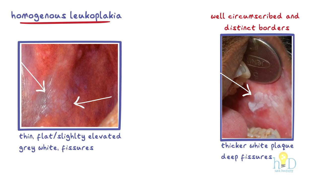
Understand not all leukoplakia lesions would look the same. There would be variations in their manifestations.
Being exposed to the different clinical variations of leukoplakia lesions would help you make a better diagnostic decision than someone who has not been exposed to as many patients as you have been. You are able to recognize patterns in the clinical variations of leukoplakia lesions.
Now consider this equation
Attending to "N" number of patients with leukoplakia = Observing clinical manifestations of "N" number of leukoplakia lesions
You see, there are only so many patients you could attend to as a student (“N” number of patients with leukoplakia). But what about the other half of the equation (observing clinical manifestations of "N" number of leukoplakia lesions)?
I used to sit in the library, and check every possible book in the Oral Pathology and Medicine section for pictures of different oral diseases. That’s it. I just looked at the pictures. I made it a point to observe pictures of one lesion every day in every possible book. I used the internet too. Just intently observed the pictures. You see, there would be slight differences in the clinical appearances of a disease in different pictures.
I was essentially artificially “simulating” a clinical scenario. Of course, there is nothing like looking at a lesion clinically, but let me tell you, this is an under-rated exercise. Observing numerous pictures of a disease intently everyday would help you develop a knack for recognizing it.
I used to literally play games. I used to open a random page, look at the picture of a lesion and try to “spot-guess” what the lesion was.
🤨Okay, but what next?
Of course, I would be wrong most of the times. But why was I wrong?
The lesion in the picture may look like another lesion -> there are several similar looking white or red lesions, for example.
This is a contrived experiment. I have no clinical details. What is the patient’s complaint? Is the patient a male or female (only oral cavity visible in the picture)? Age of the patient in the picture? Other relevant details like habits?
Apart from recognizing patterns in the clinical manifestations of different oral diseases, you are also reverse engineering and picking up on other essential details like I mentioned above.
Rather than reading how the lesion looks like and then looking at the picture, you observe the picture of the lesion, ask yourself questions (what could be the complaint of a patient manifesting this lesion, male or female, was this a result of unwanted habits, describing the clinical appearance of the lesion to yourself and so on) and then read about the clinical features of the disease. This is an active exercise, helps you connect neural dots and makes retrieval from memory easier.
🧠Retrieval of information from memory
Rather than mapping lesions from their roots (salivary gland lesions, epithelial pathology, soft tissue lesions, blood diseases etc) try mapping lesions according to how and where they could manifest. Here are a few examples:
Ulcers, red and white lesions, lumps and swellings on the lips
Ulcers, red and white lesions, lumps and swellings on the tongue
Ulcers, red and white lesions, lumps and swellings on the gingiva
Radiolucent, radiopaque and mixed radiolucent-radiopaque lesions
Make it a habit to read/map lesions this way. Everyday try recollecting different lesions according to how and where they manifest. It is easy for you to recollect, for example salivary gland lesions. However, try recollecting lesions that could appear as swellings on the palate or gingiva. This is a tougher exercise, albeit a better one.
Training yourself this way makes it easier (in the long run) to quickly list a set of diseases for differential diagnosis.
Coupling Pattern recognition ➕ Retrieval of information from memory with other details (clinical history and examination)
I’ll come back to a statement I made earlier
What if I could quickly come to a rough diagnosis of possible lesions, even before I went deep into history taking and clinical examination?
You see, repeatedly practicing the above experiments could help you make a temporary diagnostic decision quickly. I say temporary, because you haven’t elicited clinical history yet nor have you clinically examined the patient thoroughly. You look at the lesion, recognize patterns and retrieve limited information from memory (lesions manifesting as swelling in the gingiva or palate etc).
This helps you not get overwhelmed. You are calm. You have a sense of what you are looking at. Having a rough idea of what you may be looking at, gives you confidence and you ask the right questions to the patient.
Next, perform a thorough and detailed history taking and clinical examination -> arrive at a diagnosis!
Cheers
Dr.Sanketh from HackDentistry
If you want to take a deep dive, log in to our website www.hackdentistry.com
(psst, be sure to bookmark it, just in case) to read and learn with our course bundles!


