Junctional Epithelium - Histology
What is the Junctional Epithelium?
The reduced enamel epithelium is a thin membrane of cells wrapping the entire enamel surface of the tooth.
It is a layer of flat cuboidal cells and is formed by fusion of ameloblast layer and the outer enamel epithelium.
As the tooth starts to move upward and erupts through the oral mucosa, the reduced enamel epithelium fuses with the overlying oral epithelium to form the junctional epithelium or the attachment epithelium.
🎥HackDentistry - YouTube
A quick shout out! Here's a “video version” of the article.
Junctional Epithelium - Histology
The junctional epithelium is a stratified non-keratinized epithelium, 15–30 cells thick coronally and around 3-4 cell layers thick apically.
Like, rest of the oral epithelium, the junctional epithelium too, keeps proliferating in the deep layers and moves up layers to replace cells that are shed.
The cells of the junctional epithelium have a high turnover with cells continuously being regenerated every 5-6 days.
But, unlike oral epithelium, cells in all the layers of the junctional epithelium are incompletely differentiated.
They also possess lesser number of tonofilaments and desmosomes.
Internal and External Basal lamina
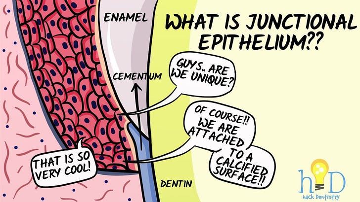
Junctional epithelium is attached to the tooth surface by means of an internal basal lamina, which is composed of cells of the junctional epithelium attaching to the tooth with a hemi-desmosome.
This is similar to the hemi-desmosomal junction between cells and connective tissue elsewhere in the body, but this junction is unique as cells here adhere to a calcified surface.
It also has an external basal lamina, where the cells in the stratum basale of the junctional epithelium attach to the underlying connective tissue with hemi-desmosomes.


