
Structure of Alveolar bone (Note & Video)

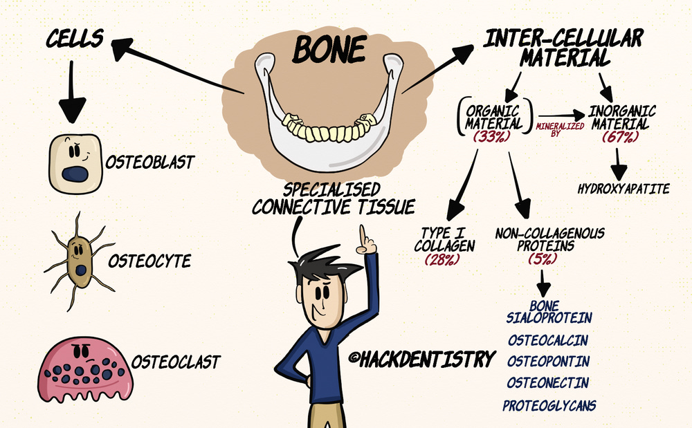
This video covers both the basics of bone histology and the structure of the alveolar bone. However, since this topic can be overwhelming I have split the whole video topic into two notes - one covering bone histology and the other covering the structure of the alveolar bone. This note covers the structure of alveolar bone.Bone is a specialized form of connective tissue that consists of cells and intercellular material.
Osteoblasts, osteocytes and osteoclasts make up the cells of the bone, while the inter-cellular material is made of organic and inorganic material.
By dry weight, bone comprises of 33% organic matrix, with 28% representing type I collagen, and non-collagenous proteins like bone sialoprotein, osteocalcin, osteopontin, osteonectin and proteoglycans making up 5%.
The organic matrix is mineralized by hydroxyapatite (inorganic material), which comprises the rest of the 67% of bone by dry weight.
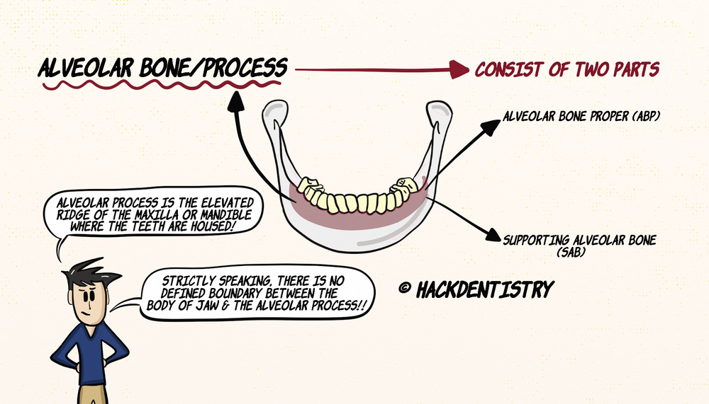
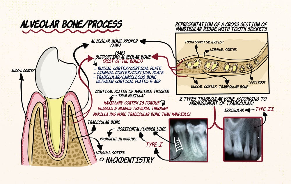
ALVEOLAR BONE/PROCESS
The alveolar bone also called the alveolar process is the elevated ridge of the maxilla and the mandible where the teeth are housed.
Strictly speaking, there is no defined boundary between the body of the jaws and the alveolar process.
The alveolar bone or process can be divided into:
an alveolar bone proper and
a supporting alveolar bone.
Alveolar Bone Proper
The alveolar bone proper (ABP) is the bone forming the tooth socket or the alveolus that surrounds the roots of tooth.
Histologically it is a compact bone having osteons.
It is also called bundle bone since bundles of principle periodontal ligament fibres called Sharpey’s fibres attach to the bone from the tooth.
The ABP or the alveolar sockets have numerous perforations of small canals that carry nerves and blood vessels. Hence the ABP is also called cribriform plate.
The alveolar sockets are separated by an interdental septum and have inter-radicular septa separating roots of the tooth.
The interdental and the inter-radicular septa are also cribriform plates and house numerous canals called canals of Zuckerkandl and Hirshfeld.
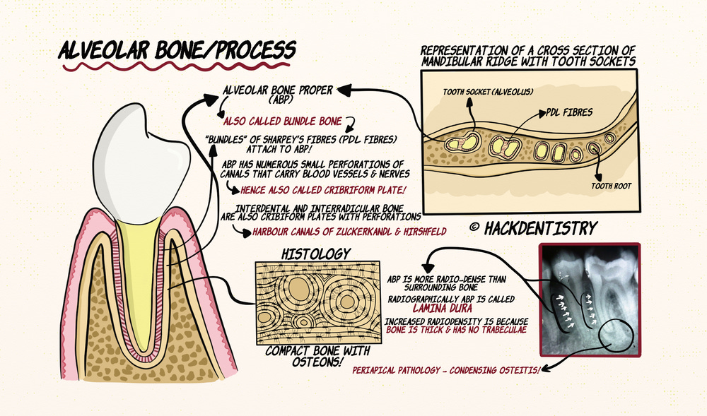
Note
In radiographs, the ABP is more radio-dense than the surrounding bone and can be seen as thin radio-opaque bone surrounding the roots of teeth. Radiographically, the ABP is called lamina dura. Its increased radio density is not because of high mineral content but due to the bone being thick and not having trabeculations.
Supporting Alveolar Bone
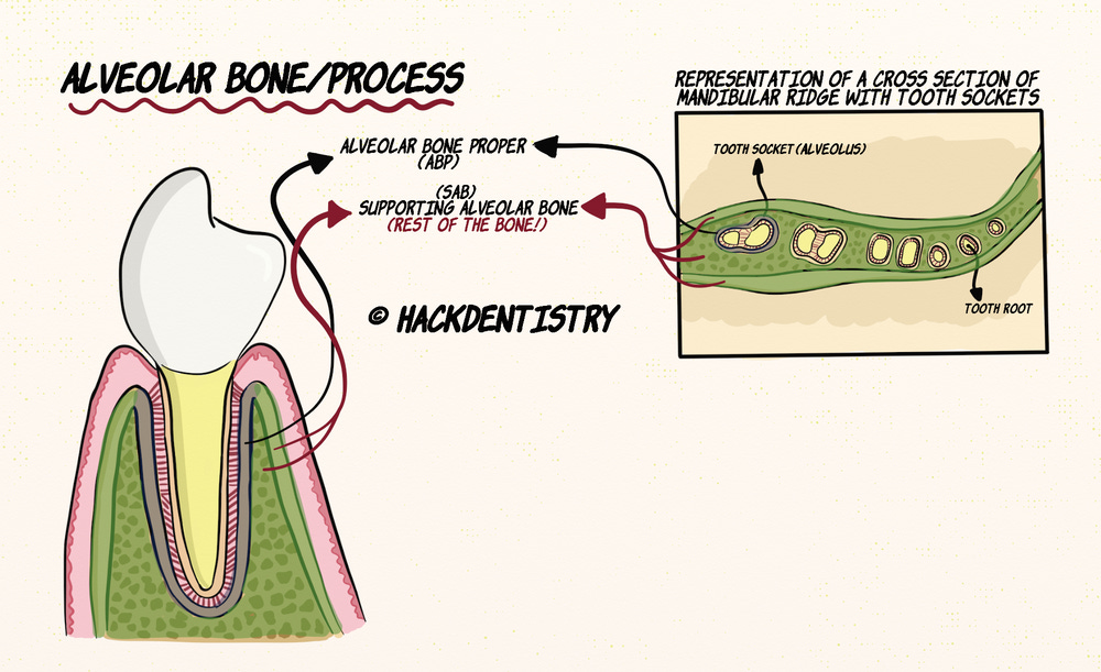
The rest of the alveolar process gives support to the ABP and is called supporting alveolar bone (SAB).
The SAB consists of the buccal and lingual cortical plates and trabecular or cancellous bone in between the cortical plates as well as the ABP.
Cortical and Trabecular bone in the Alveolar Process
The cortical plates of the mandible are thicker than the maxilla since maxillary cortex has numerous pores where many blood vessels and nerves traverse.
Also the maxilla has more trabecular bone than the mandible.
The trabecular bone according to its arrangement in the alveolar process is divided into two types called type I and type II.
Type I trabecular bone has a horizontal or a ladder like arrangement and is usually more prominent in the mandible.
Whereas the type II arrangement is an irregularly arranged trabecular bone and is more common in the maxilla.
HIGHLIGHTS - VIVA & ENTRANCE EXAM PERSPECTIVE
Osteoblasts, osteocytes and osteoclasts make up the cells of the bone, while the inter-cellular material is made of organic and inorganic material.
The alveolar bone also called the alveolar process is the elevated ridge of the maxilla and the mandible where the teeth are housed.
The alveolar bone or process can be divided into:
an alveolar bone proper and
a supporting alveolar bone.
The alveolar bone proper (ABP) is the bone forming the tooth socket or the alveolus that surrounds the roots of tooth.
ABP is also called bundle bone since bundles of principle periodontal ligament fibres called Sharpey’s fibres attach to the bone from the tooth.
ABP is also called cribriform plate.
The interdental and the inter-radicular septa are also cribriform plates and house numerous canals called canals of Zuckerkandl and Hirshfeld.
Radiographically, the ABP is called lamina dura.
The rest of the alveolar process gives support to the ABP and is called supporting alveolar bone (SAB).
The trabecular bone according to its arrangement in the alveolar process is divided into two types called type I and type II.
REFERENCES AND FURTHER READING
Nanci A. Tencate’s Oral Histology. Development, Structure and Function. 8th ed. Elsevier; 2013.
Young B, Woodford P, O’Dowd G. Wheater’s Functional Histology. A Text and Colour Atlas. 6th ed. Elsevier Churchill Livingstone; 2014.
Kumar GS. Orban’s Oral Histology and Embryology.13th ed. Elsevier; 2011.
Lindhe J, Lang NK, Karring T. Clinical periodontology and implant dentistry. 5th ed. Blackwell Munksgaard;2008.